It's common for women to have a 7 week ultrasound and for many, it's their first real look at their baby. A dating scan at 7 weeks helps to confirm baby's presence and development, but it can also help to rule out pregnancy complications.
At , the embryo matures to a foetus and individual genetics and growth factors begin to influence growth. An early pregnancy dating scan, when compared with scans later in pregnancy, can be more accurate when assessing the expected date of delivery. With maturity, the size of the baby correlates less to its age than in the early weeks
- To see if one or more babies are present and assess gestational age. A 7 week scan is sometimes called a dating scan, because it's an accurate way of assessing a baby's age and growth.
- If you've had any complications such as blood loss, an ultrasound could help identify the cause and source of the bleeding.
- To confirm the presence of a heartbeat.
- To do a general check of your uterus, fallopian tubes, ovaries and pelvic organs.
- To ensure the embryo has implanted within your uterus and is not growing outside uterus e.g. an ectopic pregnancy.
- If there's uncertainty about the date of your last normal menstrual period (LNMP). Also to assess the embryo's growth in relation to your menstrual history.
When is a dating scan necessary?
- For women who have an irregular menstrual history or cycles, an ultrasound scan at 7 weeks is often recommended.
- For women who have recently had a miscarriage and conceived again quickly. Women, who have had fertility assistance or a history of obstetric problems, are generally keen for as much reassurance as possible.
- For women who have conceived after recently stopping contraception.
- For women who are breastfeeding and have conceived again even though they may not have resumed menstruation.
- In other situations where confirming the gestational age of the baby is important.
How big will my baby be at the 7 week ultrasound?
Your embryo will be measured from the top of its head, the crown to its bottom or rump . This is called the crown-rump-length or CRL. This is because it is the longest portion of the baby's body gives an ideal measurement of its growth and development.
An average length of the embryo at 7 weeks is between 5-9 mm long. The average weight is less than 1 gram. Obviously, every pregnancy is unique and individual factors influence the size of the embryo.
How will my 7 week scan be done?
There are two ways of having a seven week ultrasound. One is via the abdomen transabdominally and the other is through the vagina transvaginally. At 7 weeks gestation, transvaginal ultrasound provides the best and most accurate visualisation. This is because the transducer does not need to send sound waves through multiple layers of muscle and tissue to pick up the returning images.
When a transvaginal ultrasound is done at seven weeks gestation, the transducer is placed in the mother's vagina and the sound waves are transmitted via her cervix directly into the uterus. With this form of scan it is not as necessary for the mother to have a full bladder. When having a transabdominal ultrasound, however, a partially full bladder is recommended. This helps to move the bowel out from the pelvis into the abdomen so the uterus, ovaries and pregnancy can be seen more clearly. Later on as the pregnancy progresses, a full bladder is not necessary as the enlarging uterus is no longer contained in the pelvic rim.
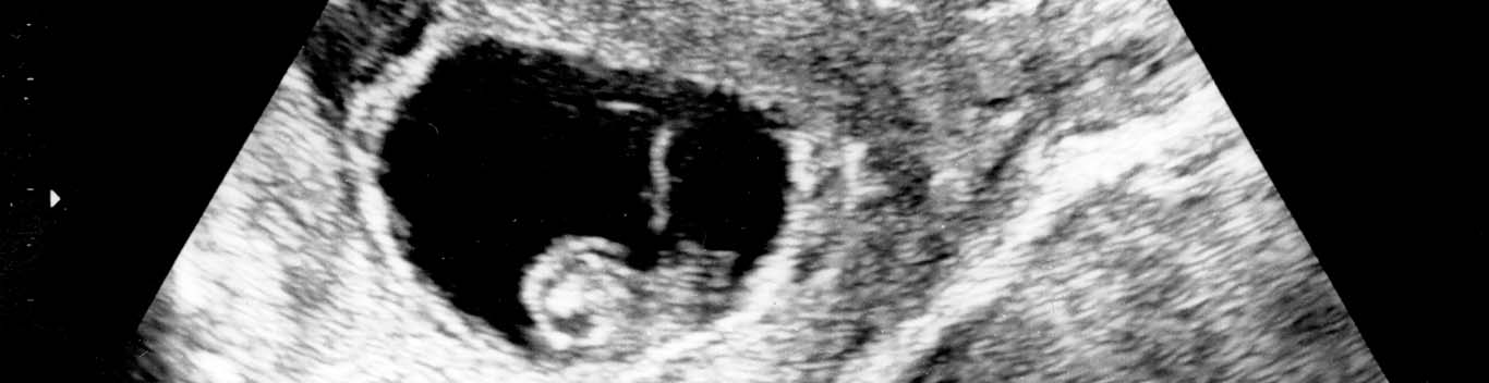
But I can t see a thing!
In very early pregnancy, the embryo and pregnancy sac may simply be too small to see very much at all. It's the shape and general structures which are more obvious. And if present, a tiny heart beat. The average number of beats per minute (BPM) is between 100-180 between 6-7 weeks of gestation.
Am I having a boy or a girl?
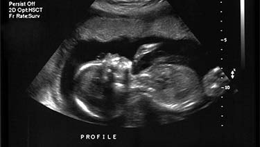
At seven weeks of gestation it is still too early to identify what gender (sex) the baby will be. It is also impossible to do a thorough foetal screening assessment because it is still just too premature in terms of embryonic development. However, general mass structures such as a head and body can generally be seen in the embryo at seven weeks.
Questions sonographer may ask at your 7 week scan
- When was your last normal period, and is your cycle regular?
- How did you conceive naturally or with fertility assistance?
- What number pregnancy this is and your obstetric history.
- How many (living) children you have.
- Your medical and surgical history.
- If you are taking any medications.
- If you've had any miscarriages or pregnancy complications.
Written and reviewed by Jane Barry, midwife and child health nurse on 6/01/20.
FAQ
Yes, one of the primary reasons for a dating scan at 7 weeks is to count how many gestational sacs and embryos are present.
If possible, this is a nice experience to share. For many partners, this is the first time the pregnancy seems real .
At the 7 weeks can, only a gestational sac and yolk sac may be seen. It's still very early in the pregnancy. If there are concerns, you may be asked to return for another scan in 7-10 days to check on the embryo's development.


Last Published* May, 2024
*Please note that the published date may not be the same as the date that the content was created and that information above may have changed since.